Introduction
General description of the spinal cord
The spinal cord forms one of the important organs of the central nervous system. It coordinates all the body functions including the functioning of various body parts and coordination between the brain and the respective body parts. In other words, it is through the spinal cord that various body signals are transmitted right away from the medulla oblongata of the brain to various body parts for a response.
Therefore any damage to the spinal cord might mean paralysis of various body parts especially those located below the area where the spinal cord is injured and this is due to failure to receive signals or coordination from the brain (Karki, 2002).
The problem
Spinal cord repair is one of the most currently exciting frontiers in the field of medicine especially the traumatic based injuries of the spinal cord. Although recent developments in the treatment sector are trying to help many people survive from injury to the spinal cord, most cases of injury to the spinal cord still pose a challenge to many other victims by leading to lifelong disability thus calling for a more critical and continued research (Zhou et al., 2003).
Since the spinal cord is a complex organ that has various cell types including nerve cells, nerve fibers connecting it to the brain and other supporting cells, the main problem that occurs when the spinal cord is injured is that these cells die and the nerve fibers lose communication to the brain thus leading to a body coordination and sensation crisis. The main challenge to the treatment measures is how these cells can be regenerated and how the fibers can be amended to regain normal functions. Since the spinal cord is uniquely arranged in lower and upper segments with the lower segment coordinating the body’s lower parts and the upper segments taking control of the upper body parts, any damage to the spinal cord poses a threat to disorganizations and dislocation of these segments whose return to fit the appropriate organization once disorganized is very complicated to the medical professionals (Ramon et al., 1998).
Though the spinal cord gets injured when subjected to trauma or accident, what determines the recovery process, the time period that the spine will take to heal and the treatment prescriptions that will be given is the severity of the injury. An injury to the spinal cord is considered severe when the pathways of the nerve fibers are badly damaged and also when there is evidence of secondary damage after the initial damage. There are various types of injuries to the spinal cord and each type will also determine the severity of the injury and the medical prescriptions. Following the injury, some patients become paralyzed due to loss of function by the nerve fibers thus no sensation and transmission of neuronal signals. Other patients may start to develop complications like pressure scores among other diseases of the respiratory nature (Bregman et al., 2002).
The medical field has been awake doing everything possible to save the lives of patients with spinal cord injuries and at the same time, researchers have been alert to develop advancements in the way spinal cord injuries can be handled. With their research, they have been able to come up with drugs therapies that are given a few hours after the patient suffers from the injury and they have been able to do imaging of the spinal cord to assess the damage. These are just some of the advancements in easing the handling of patients with spine injuries.
The acute situations of spinal cord injury are currently managed by first diagnosing the damage then trying to relieve the misalignments of the spinal cord segments that might have been diagnosed among other structural misalignments of the spine. Sometimes the occurrence of secondary damage is obvious in specific cases and the medical attendants can only minimize it by ensuring that the vertebra is stable apart from minimizing cellular damage. After the stabilization of the patient, long-term recovery is achieved by supportive care among other rehabilitation strategies. Most patients who suffer spinal cord injury become traumatized for fear of the possible outcome. This only does more have to them by escalating the injury level by means of secondary damage and this is the major challenge to efforts made to correct injuries to spinal cords. It is researched that the immune cells of the body rarely enter the spinal cord. After the injury, these immune cells enter the spinal cord and engulf the debris and eventually release powerful regulatory chemicals (Ramon et al., 1998).
These chemicals are both of benefit and also cause harmful effects. The purpose of these immune cells is still under research as very little is known about their role after the spinal cord has been injured. Antioxidants are also released after the injury and these antioxidants are free radicals and they attack the immune system. The damage to axons which are nerve fibers that passes signals to other cells is the main cause of spinal cord problems after injury. There was an assumption that these axons are damaged during the injury due to physical forces involved but the new evidence suggests that these axons slowly deteriorate after the injury due to disruption of the transport of essential molecules to and from the ends of the axon (Houle et al., 1992).
After the injury, there is the likelihood of death of the nerve cells located below the spot or lesion of injury thus disrupting the movement of signals and sensory information. The only tool to solve these entire problems is first understanding the changes that occur following the injury after which an appropriate recovery measure is applied (Zhou et al., 2003).
Cells and tissues that repair
After the injury to the spinal cord, both the nerve cells and those cells responsible for the provision of myelin to aid in appropriate properties of conduction are lost. Therefore there is a need to replace these cells and the cells that are used to repair these cells are the ones we are referring to as cells that repair. There are many cells and tissues that have been incorporated into the injured spinal cord to restore the functions that might have been lost during the injury. The cells that are used to repair include the stem cells, the sheathing olfactory cells which are those cells that perform a function of ensuring that myelin is formed on the olfactory nerves. The Schwann cells are also used as repair cells and they function to ensure that myelin is formed on the peripheral nerves (Lankford et al., 2002).
The work of these cells and tissues is to ensure that the adult neurons which might have suffered the injury or damage are provided with regenerative pathways for recovery. They also play a role in the direct replacement of the lost neurons or simply rescuing them. The main function of cells and tissue that repair the injured spinal cord is to spear the integration and regeneration process so that the spine can regain its functionality as normal (Bregman, 1988).
Due to the persistence of nerve fibers as demyelinated fibers, most of the efforts done to restore or replace damaged cells are geared towards getting cells that can give myelin to restore back the lost conduction of signals and sensations and these are the type of cells that are recommended to be transplanted. Olfactory ensheathing cells, as well as stem cells, have proved to be viable in spinal cord repair from the experiments done. The Schwann cells and oligodendrocytes worked in some cases and failed in other cases. There is great hope in the future due to the ability to alter the secretory characteristics of the cells to be transplanted meaning that many more alterations can be made to make the transplanted cells compatible with the patient. These alterations in most cases employ the use of molecular techniques which can allow the addition or removal of one or several factors with different significances to the recipient like chronic pain reduction (Lankford et al., 2002).
The idea of bioengineering of cells by use of gene therapy is beneficial in a way that it employs the use of two factors and combines them to give a more sound and pronounced outcome. The product of a combination of the gene delivery system and cell’s therapeutic advantage gives bioengineered cells the following desirable characteristics. The resultant cells are considered vital due to their adaptability and compatibility with the hosts in which they are transplanted for purposes of spine repair. The best demonstration of these bioengineering combinations is in a case where the patient suffering spinal cord injury requires to be delivered with neurotrophin. In this case the there can be bioengineering of two cells to come up with this desired combination. One cell that is capable of producing myelin will be bioengineered with another cell that is capable of secreting neurotrophins. This combination will be able to give rise to a cell that can produce myelin to assist in the restoration of the functions of the spinal cord and at the same time enhance neurite growth in the injured spinal cord (Bracken et al., 1992).
Taking an example of attractive populations of cells meant for studies concerning cell transplantation like fibroblasts, its main strength is that it can make the patient harvest his or her own fibroblasts including culturing of the fibroblast of the patient with very fast proliferation rates shown in vitro and lastly its ability to access to transfection with ant gene that is desired in by the patient for transplantation. This cell transfection was tried in rodents and the results came out positive showing the stated abilities of fibroblast when transplanted but the only challenge that was realized was that these cells produced collagen which we know is not normal for it to be found in the spinal cord. Apart from this challenge, the ideas helped in the targeting of other cells and genes that can be harvested from respective patients by the use of gene therapy. Cell transfection by use of neurotrophins has been proved to work well with embryonic precursors as well as Schwann cells. The two have shown recovery of both the behavior and also neurite growth has been promoted (Finney, 1993).
In addition to replacement cells and tissues, the endogenous cell population of the injured spinal cord also needs to be boosted and this is only done by macrophages which are activated by their own patient. The macrophages are normally found in the blood and when a person is injured like in this case where the spinal cord is injured, these macrophages are supposed to be activated and transferred to the area where the injury has occurred and on arrival, they trigger the growth of cells present at the site of injury thus enhance faster healing of the injured spinal cord or any other part that might have suffered an injury.
The advantage with macrophages that are activated by the patient himself or herself is that they are capable of scavenging the myelin debris that might already have started degenerating and since they are composed of nonpermissive factors, they immediately trigger the regenerative growth process without necessarily triggering a response by the immune system. The discussion of the cells and tissues that repair the damaged spinal cord as demonstrated above aims at achieving either of the following. The repair cells should at least be able to replace the damaged cells directly or repair the existing cells by stimulating the production of some growth-promoting conditions to enhance patient recovery (Finney, 1993).
Inhibitory factors
The main action of the inhibitory factors is to hinder the regeneration process by preventing the re-growth of axon fibers and other nerve cells. The makeup of the neurons found in the CNS which are highly susceptible to trauma also affects the regeneration process. In this discussion, the focus will be on inhibitory factors.
Myelin inhibitors are one of the inhibitory factors of focus. They are protein fractions that are found lying in the central nervous systems. This protein has been identified as one of the first factors that cause inhibition of growth of the neuritis of the central nervous system. Currently, research has given it a new name and it is termed Nogo-A. As per the experiments done, the Nogo-A prevents the outgrowth of neurites in vitro while in vivo, studies have shown that the neutralization of the Nogo-A functionality indeed showed recovery of the functional properties, plasticity was also recovered and, more so, the regeneration of the neurites or the axons was also among the observations made (GrandPre et al., 2002).
From these experiments, it is now clear evidence that indeed the myelin protein fraction of the central nervous system also known as the Nogo-A protein fractions is actually an inhibitor to the growth of the neurites although some of the experimental outcomes are still controversial. Other growth inhibitors associated with myelin include the following. One of them is the glycoprotein associated with the myelin of the central nervous system. This glycoprotein is given a code as MAG108 and its characteristics as an inhibitory agent or factors have been too proved from various Vivo experiments where its effect has been blocked or inactivated thus giving results of effective regeneration of the axons or neurites of the central nervous system. Among other observations made include recovery of the normal functions of the injured spinal cord and plasticity recovery (Jones et al., 2003).
Actually, these results are the same as the one obtained when the Nogo-A inhibitor was inactivated in an experiment to determine if it is actually a growth inhibitor. Another myelin-associated glycoprotein called oligodendrocyte coded as OMgp also showed growth inhibitory characteristics just like Nogo-A and MAG108. The problem is that there is still more to be covered involving the inhibitory characteristics of the above-mentioned myelin-associated proteins and the effect of neutralization of their activity is also yet to be explored.
Chondroitin sulfate proteoglycans are also another inhibitory factor. The general description of proteoglycans is that they are simply forms of molecules of the matrix that are extracellular in nature. The molecules are composed of a protein core with moieties of sugar molecules denoted as GAGs attached. GAGs mean glycosaminoglycans. There are other complex chemical constituents associated with this proteoglycan all found within the central nervous system and they are all associated with causing inhibition to the growth and regeneration of the axons and other dells of the spinal cord that diminish after injury to the spinal cord. These components cause inhibition of neurite growth. The results obtained in vivo are that the components they too inhibit the growth of the neurites. These were also proved from various sets of experiments that saw the effect of these inhibitory chemical components blocked or neutralized thus giving outcomes as follows. The observations were that the regeneration of the axons and neurites was evident and that the functionality of the spinal cord was recovered. The only thing remaining as far as research is concerned is to establish the effect of neutralizing these inhibitory factors (Simonen et al., 2003).
There are also other minor inhibitory factors that are capable of preventing the growth of the neuritis and axons among other recovery procedures like recovery of the functional effectiveness after injury of the spinal cord and also plasticity recovery. Semaphorins and ephrins among other growth limiting factors are some of the examples given (Merkler et al., 2001).
Scar
The definition of scar is that it is the tissue that forms alongside inflammation of the injured spot on the spine. This tissue is normally comprised of sugar protein that inhibits the regeneration process of the injured spinal cord.
Bioengineered regeneration strategies of the spinal cord injury
There is no other strategy that can be applied after the spinal cord has been injured in order for it to regain its normality or successful regeneration apart from ensuring that the nerve cells that were injured during the spinal cord injury or did not survive the injury are replaced and that the axon fibers that were damaged find their way to re-grow so that they can resume their normal functions of signal transmission and sensation by finding their appropriate targets.
Otherwise, all the other efforts will be null and void or will just be a supplement to the above. The main aim of regeneration strategies is to ensure that the disruption on how the segments of the spinal cord are arranged is brought back to normal so that the axons match their specific targets for successful passing of the signal and by doing so, the normal functions of the spine are likely to be restored This process is complicated and poses a great challenge to the biotechnology field of medicine. Failure of the axons to interact with their specific targets to come up with a synopsis is what is described as paralysis.
The requirement for the regeneration of the injured spine matches those required by the developing spine and this has been the main focus of research. The research is based on finding out the type of cells that are responsible for the development of the spinal cord, how these particular cells undergo differentiation and specialization during the spinal cord development process and how the axons of the developing spinal cord get to their respective targets in order to create appropriate synopsis in the spinal cord. All this is done on the uninjured spinal cord so that the findings can be applied to the already injured spinal cord. Based on the several biotechnological types of research done on the regeneration of the spinal cord, the neurons of the CNS can only be able to resume growth after a combination of specific chemicals is attained. These chemicals are natural in nature and are termed trophic factors. Thereafter, the bioengineered strategies that are applied to correct injuries on the spinal cord and create a permissive environment for the regeneration process all have based on identification and understanding of the specific trophic factors that can be biotechnologically combined and induced to the patients to commence the process of regeneration of the spinal cord. However, there are risks of side effects of these strategies and therefore each one of them needs to be examined first for such risks of side effects before they are typically applied in the cases of human beings.
The innate ability of nerve cells to grow is currently on watch by researchers after which they will find out the appropriate environment for their growth and by doing so, they can apply these conditions in the biotechnology lab to get growing nerve cells and induce them to patients after successful experiments so that the nerve cells can induce the regeneration process of the injured spinal cord (Mitler 2003). Application in animal models of these strategies has also been done by researchers.
Among these strategies include grafting of peripheral nerve pieces and fetal tissue into the spinal cord that has suffered damage or injuries,
administering of the necessary and appropriate growth factors,
manipulation of the programs of cell death genetically, and neutralizing or inactivating substances that naturally inhibit the growth of the cells.
Combinations of such therapies have enabled the coming up of the first evidence that there is a great possibility of some functional regeneration occurrence in injured spinal cords including some completely severed spinal cords in grown-up species of mammals (Andrews, 1986, p. 132).
Repair by cells.
Transplantation
Cell transplantation is one of the bioengineered strategies that can be applied to correct injury to the spinal cord. There are many trials in place to find out the viability of these transplantations. The projections are that some will work while others will not. To be more specific, the kinds of cells that are being applied in this technology are those that are able to create an environmental situation that will permissively allow the growth of axons. If not cells, then the peripheral nerve pieces. These cells or peripheral nerve pieces have the ability to generate some growth-enhancing substances to the axons and other nerve cells (Levi et al., 2002).
The creation of such conditions is also ideally proved to enhance the marching of the neural nerves to their respective and appropriate targets and this helps in quick recovery of the normal functioning of the spinal cord. When these PNS are transplanted, there is less scarring observed around the area where the injury occurred sine the scarring effect may impede the re-growing of the axons. The best example of cell transplant is where the technique was tested in rats (Bomstein et al., 2003).
In this case, the Schwann cells were the cells that were used in the transplant experiment. “The Schwann cells are actually forms of glial cells and they are diagnosed with the ability to promote the growth of PNS axons” (Hofstetter et al 2002, p. 203). These cells were injected into the injured spinal cord of rats. The control experiment was also done where the rats with injuries on spinal cords were not injected with this cell transplant. The results of the experiment were that the rats that were injected with the cells showed evidential re-growth of the axons and the damaged nerve cells while those that were not injected with the cell transplant did not show any signs of axon regenerations and repair of the damaged nerve cells. The technique is currently under trial test where the human Schwann cells are still under study to determine if the same can be applied in cases of human injury to the spinal cord can be corrected by similar cell transplantation (Hofstetter et al., 2002).
The other transplantation biotechnology is the implantation of fetal tissue. The fetal tissue is studied as a potential enabler of the axon regeneration process. These tissues obtain these characteristics from the fact that it contains many substances capable of supporting growth among them being the stem cells and progenitor cells. The tissue has also proved to provide a conducive environment for axon growth with fewer obstacles involved if any (Nieto, 2003).
Due to the ability of the stem cell to differentiate and form several cell types, the transplantation of these tissues that indeed contain these stem cells may lead to the development of supporting cells or neurons in the spinal cord and this will have contributed a lot to the recovery of the injury but this will only happen if the appropriate signal is received by the stem cells (Tessler, 1991, p. 118).
This research has also been experimentally proved in the case of rats just like the Schwann cell above and the results were that the rats implanted with these tissues showed progressive recovery from the spinal cord injury and there was observable re-growth of the axons or axon regenerations at the injured sport. However, no axon regeneration or growth was observed in the case where the rats received no transplant. This shows the effectiveness of these fatal tissues (Finney, 1995).
Stem cells
The appropriate definition of stem cells is that differentiate along lineage pathways to form other types of cells and the types of cells that result are usually not the same as the initial type of the stem cell used. This phenomenon is termed self-regeneration. The stem cells that treat spinal cord injury must have the following basic characteristics. They must be able to gather and be found in very large amounts like in billions or several millions of them. The cells must be able to permit harvesting by an invasive procedure that is minimal. They must also meet the above definition by having the ability to self regenerate along lineage pathways. The other factors that need to be met by these cells include their capacity to be manufactured with the good observation of the rules and regulations applied for transplant cells’ bioengineering. “The cells must also be able to be transplanted safely as well as effectively to a host living thing that is either allogeneic or autologous” (Herman et al., 2002, p. 66).
Stem cell therapy
Stem cell therapy relies on one characteristic of stem cells and this feature involves the ability of the stem cells to undergo the regeneration process and result in a number of cell types different from the initial type with the embryo, the brain, and the borne marrow being the major sources of these cells. There are different types of stem cells that are applied under this therapy. The neural stem cells when used usually help in the generation of neurons as well as glia. These cells are currently under research in animals like rats and hippopotamus with the hope that the same will be applied in human beings. When demonstrated in rats, the cells have shown differentiation into different types of cells among them being the astrocytes and oligodendrocytes but the recovery implication is yet to be established.
The experiment that involved transplantation of the human neural stem cells to adult rats which had a spinal cord injury showed some positive results where the axon myelination, as well as electrical conduction, was nearly the same as that normally in-process and eventually there was some recovery exhibited by observations of some form of axon regeneration of the spinal cord (Myckatyn et al., 2004, p. 354).
When the same experiment was done using the hippocampus as a host, the cells that were injected were closely monitored and gave the following results. After some period of monitoring, it was realized that the cells had migrated to the injury spot and on arrival, they induced an observable regeneration of the axon fibers. The conclusions were that there is a possibility of the same effect o happen in human beings host with further trials.
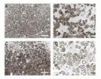
The above figure shows remyelination of the spinal cord of the rat following transplantation of adult human precursor cells. Normal (A),
demyelinated (B), and remyelinated axons (C) of the dorsal column. (D)
Remyelinated axons at higher magnification. The anatomical pattern of myelination was similar to that produced by Schwann cells (arrows). (Bar:
A–C, 25 µm; D, 10 µm) (Akiyama et al., 2001, p.33).
Bone marrow-derived stem cells are mostly used due to their functional ability to affect the remyelination process which is a start journey to spine recovery and a number of ways are applied when administering these stem cells.
The first way is by direct administration and the second method is by intravenous application or administration. In cases where these stem cells are transplanted and fail to give rise to long axon regenerated fibers, the directly applied cells normally form neuron-filament bundles and these bundles enhance the formation of long axon fibers that is able to link the graft tissue to the host and these long axons are able to regenerate and extend the whole lesion either across or along with the lesion.
This demonstration experimented in rats where the injection of born marrow stem cells made the rats that had injuries on the spinal cord regain some locomotive activity and this relates to the future postulation that actually the same will happen when stem cells from human bone marrow are applied in human cases to correct the injuries to the spinal cords of human beings. The same experiment also showed observable regeneration of the axon fibers at the point of injury in rats that were injected with these stem cells.
Embryo-derived stem cells are also used. Although this type of bioengineered strategy to repair damaged spinal cords has raised controversial ethical concerns, they have been proved to help a lot in the treatment of injured spinal cords. The research done on this type of stem cell indicated that they have the ability to regenerate and form other cells like neurons and glial cells which function to restore the normal function of the spinal after the loss of function due to the injury to the spinal cord (Hendriks et al., 2004).
In vivo, there have been some side effects caused in experiments performed using these embryonic stem cells to correct or repair damaged stem cells thus need for more trials before effective use. If this technology is fully implemented, the medical attendants need to be very careful with the number of cells transferred of the type of stem cell therapy to be prescribed so as to avoid incidences of patients reacting negatively to the therapy or simply developing side effects.
Inhibitory factors removal
For the Nogo myelin-associated inhibitory factors, the researchers have come up with an antibody called IN-1 which has been successfully proved to be able to suppress the inhibitory effects of this inhibitory factor of limiting the growth of neurites, slowing down the regeneration process, and limiting recovery of the normal functions of the spinal cord after injury. When this antibody was induced along with the peptide antagonist receptor of the Nogo inhibitor, the results were that there was a recovery of the normal functioning of the axons in the rats that were used in the bioengineered experiments (Ribotta et al., 2004).
The myelin-associated growth inhibitors can also be suppressed by the application of the high amount of cAMP secondary messenger. In the experiment to determine whether this secondary messenger was capable of suppressing the inhibitory effects of myelin-associated growth inhibitors of the central nervous system, rats with injured spinal cords were injected with a high amount of this secondary messenger near the spot where the lesion was located entering directly to the ganglion dorsal root. The control group of rats with injured spinal cords on the other hand was injected with saline DB-cAMP messenger at the same spot just like the experimental rats. Other rats were not injected with the messenger. The results were that after some period of close monitoring and investigation, the rats injected with the DB-Camp messenger showed a higher response in terms of growth of the axonal fibers which was seen at the point of injury of the spinal cord. This growth was estimated to range from 1.5 to 6.0 millimeters. In rats injected with cAMP general messenger, there was evidence of the growth of the axon fibers but this time to a lesser extent compared with the first results of the DB-cAMP specific messenger. The cAMP general messenger showed an approximate growth of about 0.6 to 0.9 millimeters of axon fibers around the spot of the injury to the spinal cord (Hill et al., 2004).
However, the results for the third set of injured rats where injection of the secondary messenger was not done expressed no observable growth or regeneration of the axon fibers around the lesion spot where the injury occurred. This shows that the first and the second set of experiments where there was observable regeneration of the axon fibers shows that indeed the secondary messenger stated has the ability to suppress the myelin inhibitory factors and thus allow for regeneration of the axon fibers. However, the third set of results shows that since the secondary messenger that was under investigation was absent, the growth inhibitors’ effect was not suppressed thus the inhibitors were active thus leading to no growth of axon fibers. This is the main reason why there was no evident growth of the axon fibers at the point where the injury occurred. This indeed shows that the cAMP is an anti-inhibitory factor.
Other studies on how to suppress the effect of growth inhibitory conditions that come up after the injury to the spinal cord usually target the prevention of the production of antioxidants which are known as free radicals. The effects of free radicals or antioxidants are not desirable at all. The environment that is created by these chemical radicals suppresses the growth of cells at the point of injury thus slowing down the recovery efforts. Other processes that need to be closely monitored and prevented from occurring include apoptosis and inflammation. Inflammation may be accompanied by the growth of the scar tissue and all of them hinder the regeneration processes and functional recovery.
In an experiment to prove this, skin-co incubated macrophages and certain specific antigens were injected in rodents that had suffered spinal cord injury. The injection was objectively meant to control inflammation of the injured spinal cord and prevent the production of antioxidant free radicals as well as inhibit the apoptosis process (Hulsebosch, 2002, p. 243).
In the control experiment, rats with injured spinal cords were not injected with these regulatory compounds and there was probable inflammation, apoptosis, and production of free radicals of antioxidants. After some period of time of monitoring both experimental sets, the outcomes were recorded and the following analysis was made. In the experiment where rats were injected with the regulatory compounds that prevent apoptosis, inflammation, and production of free antioxidant radicals, there was observable regeneration of the axon fibers and recovery of the normal functioning of the spinal cord among other improvements meaning that the regulators suppressed the inhibitory factors of the central nervous system and created permissive growth environment for the regeneration process to occur. In the control experiment where the injured rodents had inflammation on the injured spinal cords and probable production of antioxidants free radicals as well as apoptosis, the regeneration and recovery of the normal functions of the spinal cord were not observed.
This clearly indicates that conditions like controlled inflammation, apoptosis, and production of free antioxidant radicals surely suppress the regeneration process and the process of recovery of the normal functioning of the spinal cord after injury. Therefore to control such effects, it is advisable to control some conditions that may occur after the injury like apoptosis, inflammation, and production of free antioxidant radicals and this will create a permissive environment that will allow the regeneration process to be effective and see to it that the spinal cord recovers from the injury and resumes its functions (Privat et al., 1989).
Drug therapy
The application of drug therapy in cases of spinal cord injury was earlier on a great challenge to the medical officers and this only came into reality first in the year 1990 when the research on a medical prescription termed methylprednisolone successfully underwent the process of testing and other forms of clinical trials and was now allowed to be applied to other standard use purposes. This was a great breakthrough to the medical field as well as the research sector as it was the first drug therapy to be permitted to be administered in cases of spinal cord injuries. It called for great celebration and acted as a boost to the morale of researchers to even come up with more drug therapies that can successfully undergo the clinical trial procedures so that they can also be allowed to be used for standard application purposes. “The NASCIS II (National Acute Spinal Cord Injury Study II) trial” (Sadowsky et al., 2002, p. 687) which was carried out in many several medical centers to prove the effectiveness of methylprednisolone therapy was done in comparison with naloxone drug and the results were that methylprednisolone drug therapy achieved a significant improvement r recovery in humans with injuries to their spinal cords just within eight hours of application after the injury.
The results of the application of methylprednisolone showed that the patients who were completely paralyzed and given methylprednisolone were able to recover up to an average of almost twenty percent of their motor function that they had lost and compared to untreated patients only eight percent were able to recover their motor function. Paretic patients who are also termed partially paralyzed patients were able to recover an average of almost seventy-five percent of their lost motor functions compared to those who were not given this drug with a percentage of fifty-
nine percent. Patients that were administered with naloxone or administered with methylprednisolone therapy more than eight hours after the spinal cord injury actually were not able to improve significantly better than the ones given a placebo (Green, 1997, p. 541).
There was a serious revolution in terms of thinking as far as the medical field is concerned due to this successful endorsement of methylprednisolone. This successful trial opened the eyes of many medical researchers who were almost t giving up to a level of some wishing to declare that the treatment of acute spinal cord injury is impossible. This chance gave them a challenge that indeed there are still open windows for opportunities to be explored to even find many other ways in whichever spinal cord injuries can be well taken care of. In the present days, patients who suffer spinal cord traumas due to injuries to their spinal cords are prescribed by doctors with drug therapy consisting of methylprednisolone and they are normality given this therapy immediately after the injury to their spinal cords in order for it to be effective and this is normally done before the expiry of three hours just after the patient has suffered the trauma of injury and this must apply to severe cases or acute cases of spinal cord injury. Most of the medical rooms that are set to handle acute or severe cases of trauma or injury to the spinal cord are well equipped with all the requirements and prescriptions of this therapy and the patients normally receive it on arrival after suffering the injury due to awareness of the value that this drug therapy hold interns of taking care of spinal cord injury cases. The drug has been widespread and is now available n almost all parts of the world and this shows that almost all qualified health care systems put in place by governments of various countries are able to handle these cases of spinal cord injury successfully. The feasibility of this therapy as indicated by the trial made by NASCIS II shows that the therapy has very few side effects involved in a few cases if any and their work has given the body credibility to carry out such approval trials.
This is not the only drug that is expected in the future. There are several other drug therapies that are still undergoing medical examination tests or trials to prove if they can be used for standard purposes just like methylprednisolone hoping that in the future, we will be having several permitted therapies to cater to the many need of patients in terms of the pain, paralysis and other consequences they pass through after injury to their spinal cord. Among these drugs that are still underway on the testing panel include the regimen of methylprednisolone. Although this drug is almost completing the trial test, in some cases it has been administered within forty-eight hours following the spinal cord injury and it has proved to be effective. This drug therapy has been predicted to soon attain the warrant to be used as standard drug therapy in the treatment of spinal cord injuries (Ramon et al., 1998).
The GM-1 ganglioside drug therapy is also one of the drug therapies that are still in the testing process and is being tried in several clinical trials to prove its effectiveness and it is normally being tested as a measure of handling secondary damage to the already injured spinal cord since earlier on, we discussed that the injury to the spinal cord does not end at the time the spinal gets injured but rather continues due to the trauma involved and this drug is predicted to have the ability to reduce the trauma that occurs after the injury and thus help in preventing secondary damage. Other serious complications might arise in cases where the spine suffers acute or chronic injuries and this is where GM-1 ganglioside drug therapy applies most. The recovery of the neurons and the effects of the post-injury tram are also minimized by this drug. All this can either be applied at the initial treatment process or during patient rehabilitation (Ramon et al., 1998).
Scar
This is the tissue that forms at the point of injury to the spinal cord. The scar consists of the sugar proteins and this scar has been proved to impede the process of regeneration of the axon fibers and other nerve tissues (Weidner et al., 1999).
Overcoming the scarring effect
Since the sugar protein found at the scar hinders the normal process of nerve cell regeneration and re-growth, the only way to overcome these effects is to find a way of eliminating the tissue making up the scar. Research has shown that the application of an enzyme derived from bacteria species capable of digesting this scar tissue will help eliminate the scar by digesting the sugars and proteins. The only challenge that faces this mode of scar elimination is that these enzymes are usually very delicate due to high sensitivity and high degradation rate. Therefore since the enzyme is very delicate and is also thought of being very sensitive to heat, the strategy is to inject it into the body constantly for the effect to be realized (Weidner et al., 1999).
Scaffolds
Scaffolds can be simply described as a composition of synthetic or artificial biodegradable or simply biomaterials that can be seeded with appropriate neural stem cells and transplanted in the respective or desired host. The seeding of the scaffolds gives it the functionality of enabling regeneration of the axon fibers and other nerve cells. The scaffolds enable the spine to restore its lost functions and operate normally as before (McDonald, 2002).
Repair by scaffolds
When talking about scaffolds, we are talking about materials that may be synthetic or natural but have to be biodegradable in nature. They are these biodegradable materials that are seeded with progenitor cells and applied as a bioengineered strategy for injured spine repair. This type of seeded scaffold usually when applied to an injured spinal cord promotes repair of the lesions and also enables the large neural defects that might have occurred during the injury to repair. The differentiation process of the neural stem cells is also induced by these biomaterials thus enhancing recovery. These scaffolds also contain a protein known as collagen and this collagen produces collagen gel that is conducive for the proliferation of the neural stem cells. The artificial scaffolds consisting of polymers that are biodegradable some of them being PGA and PLA alongside their respective copolymers have exhibited the potential to induce spinal cord repair when combined with the neural stem cells for transplantation. These combinations were tried or experimented with in mouse and the results were that the mouse which had a spinal cord injury showed some recovery with the regeneration of the axon fibers evident. The embryonic stem cell obtained from humans also showed differentiation when applied to PLGA scaffolds and the experimental research on rats proved the effectiveness of these scaffolds (Coumans et al., 2001).
Drugs
Clinical management and drug delivery
The clinical management of injury to the spinal cord is no longer a fatal issue following the recent advancements in the biomedical field. In the last twenty to thirty years, the techniques applied to correct spinal cord injury include irrigation and cooling of the spinal cord. there was no essential understanding of why these strategies were being used by then but following the recent advancements in the medical field like the use of methylprednisolone drug therapy to correct acute cases of spinal cord injury as discussed above among other advancements like improvement of the procedures for diagnosis of the injuries caused has led to coming up of better as well as proper imaging techniques that are seeing those with severe injuries to their spinal cord recover (Houle et al., 1992).
There are three main primary considerations that medical officers or attendants take into account when addressing a situation of acute spinal cord injury. Checking for compression of any form to the injured spinal cord has no compromise once a patient with spine injury is received at the hospital. If detected, they immediately relieve the compression so that no further secondary damage progresses. Misalignments of the lower and upper segments making up the spine can also occur and need to be checked as fast as possible. When these misalignments are noticed, they are supposed to be corrected as first as possible by application of appropriate strategies discussed above and see to it that the misalignments are corrected before further developing into nonreversible complications. The second thing that the medical attendants ensure is that they make sure that there is no cellular level damage and if there is any, they try as much as possible to minimize it. Then finally, the medical attendants make sure that there is no further damage or secondary damage by making sure that the vertebrae are stable (Kwon et al., 2002).
Patients with spinal cord injuries are handled with a lot of care and attention. They are restricted from movements since such movements might cause more harm to the already damaged spinal cord. Thus the medical personnel are normally keen and treat these cases with an emergency.
Methylprednisolone is a steroid drug and is normally prescribed in acute cases of spine injury. This drug therapy functions by making sure that the cellular membranes are not subjected to injury since when the cellular membranes are left to die, definitely the neurons will also die following the injury. The drug also helps to relieve or reduce the inflammation around the spot of injury and since activation of the immune cell at the point of injury is also not desired as it enhances neuronal damage, the drug also helps to prevent this natural activation of the immune cells. This helps to spare some of the nerve fibers that would otherwise have been lost thus contributing to patient recovery (Bradbury et al., 2002).
Surgery is also sometimes an option especially when relieving the compression of the spinal cord is not achieved by other methods. Earlier on, medical attendants specifically the surgeons were very reluctant and conservative in opting for surgery since the surgeries carried earlier on proved to cause even more complications to some patients. However, the advancements in the medical field have given the medical attendants more confidence and they have been carrying out successful surgeries after x-ray examination or diagnosis with few complications involved. It is normally done as early as possible to enable early physical therapy and this prevents further complications that may likely arise following the injury and increase the probability of the patient recovering all the normal functions of the spinal cord. There are also other imaging technologies that can be applied among them being the use of computed tomography scans. These computed tomography scans are applied to be able to come up with scans with visual the nature and extend of any fractures that might have occurred on the spinal cord. Magnetic resonance imaging denoted as MRI can also be applied to image contusions among other forms of damages and all this will help in the administration of the appropriate strategy for spinal cord repair. When the spinal cord is injured to an extend where it needs support, metal plates, crews, and other appropriate devices can be inserted through surgery to stabilize or offer support (Jin et al., 2002).
As the condition of the patient continues to stabilize, the management of the condition shifts to strategies of rehabilitation and provision of supportive care but this has to be done until the patient continues with the prescribed medication. In the case where the patient suffered severe injuries and movement is a problem, changing the position that the patient is lying from among other supportive care will help a lot to prevent the development of other complications like pressure scores among other complications of respiratory nature that may attack the patient.
Physical and emotional recovery also needs to be emphasized when planning for rehabilitation measures. The muscle and other components of the injured spinal cord need not be left dormant or inactive for long time. The flexibility of the spinal cord needs to be enhanced by appropriate physical therapy and this will relieve stiffness in the joints that link the segments of the spinal cord. Otherwise, if they are left dormant, it might lead to permanent dormancy thus permanent paralysis. Physical therapy may also activate the growth of the cells and this contributes to recovery and also the risk of clotting of blood around the area of injury is also reduced. Counseling will relieve the patient emotionally and suppress the effect of trauma that the patient might have suffered during the injury (Sipski, 2003).
Summary
Although many people still believe that complications related to the spinal cord are untreatable, the medical field is putting in a lot of effort to prove this allegation wrong. The CNS tissue can still regenerate or be replaced given the evolving technology of bioengineering and provision of necessary growth conditions. Although there exist various obstacles that hinder the growth of axon fibers and other nerve cells called inhibitory factors, research has gone deep to come up with strategies in which these inhibitory factors can be blocked from their action.
The main focus when dealing with spinal cord repair is to ensure that there is the regeneration of neurons and this is only achieved under some conditions or factors. One of these factors includes ensuring the “Control of reactive astrocyte secretions, blockade of myelin-associated proteins, the introduction of neurotrophins, and transplantation of special peripheral nerve and stem cells” (Blesch, 2003, p.412). Since these conditions have been proved to create a friendly and conducive environment that permits the growth of the nerve cells with minimal obstacles involved thus promoting the regeneration process.
However, there is an urgent need for advanced research that will promote biotechnologically based therapies including those that will see to it that there is minimal host to graft interference especially when a cell of tissue transplants are being made.
The compatibility and the reception of the transplanted cells or to tissues will spearhead the regeneration process. Once this is in place, the restoration of the normal function of the spinal cord, locomotor function, and other many lost physiological functions will be achieved. The precaution however should be that any therapy applied to human beings should first be proved to be safe through various trials before approval to be used in a standard way.
Since most of the research underway concerning the bioengineered strategies for repair of the damaged spinal cord has started proving to be viable and pointing towards even more advancements, the hope is that the experimental studies will be soon translated from the laboratory neuronal regeneration to the actual neuronal regeneration in the ailing patient.
Bibliography
Akiyama, Y et al., 2001. Transplantation of clonal neural precursor cells derived from adult human brain establishes functional peripheral myelin in the rat spinal cord, Exp Neurology, 167, pp. 27-39.
Akiyama Y et al., 2002. Remyelination of the spinal cord following intravenous delivery of bone marrow cells, Glia, 39, pp. 229-236.
Blesch, A., 2003. Cellular GDNF delivery promotes growth of motor and dorsal column sensory axons after partial and complete spinal cord transections and induces remyelination, J Comp Neurol, 467, pp. 403-417.
Bomstein, Y et al., 2003. Features of skin-coincubated macrophages that promote recovery from spinal cord injury, J Neuroimmunology, 142, pp.10-16.
Bracken, M et al 1992, Methylprednisolone or naloxone treatment after acute spinal cord injury: 1-year follows up data, J Neurosurg, 76, pp. 23-31.
Bradbury, E et al., 2002. Chondroitinase ABC promotes functional recovery after spinal cord injury. Nature, 416, pp. 636-40.
Bregman, B., 1988. Effect of target and non-target transplants on neurons survival and axonal elongation after injury to the developing spinal cord. Prog Brain Res, 78, pp. 205-211.
Bregman, B et al., 2002. Transplants and neurotrophic factors increase regeneration and recovery of function after spinal cord injury. Prog Brain Res, 137, pp. 257- 273.
Coumans, J et al., 2001. Axonal regeneration and functional recovery after complete spinal cord transaction in rats by delayed treatment with transplants and neurotrophins. J Neuroscience, 21, pp. 9334-9344.
Andrews, L., 1986. bioengineered strategies for spinal cord repair. London: Methuen publishers.
Finney, A., 1995. Spinal cord injury repair. New York: American press.
Finney, M., 1993. Recent advances in pathophysiology and treatment of spinal cord Injury. Georgia: Jekyll Island.
Fujiwara, Y et al., 2004. Intravenously injected neural progenitor cells of transgenic rats can migrate to the injured spinal cord and differentiate into neurons, astrocytes, and oligodendrocytes. Neurosci Lett, 366, pp. 287-291.
GrandPre, S et al., 2002. Nogo-66 receptor antagonist peptide promotes axonal regeneration. Nature, 417, pp. 547-551.
Hendriks, W et al., 2004. Viral vector-mediated gene transfer of neurotrophins to promote regeneration of the injured spinal cord. Prog Brain Res, 146, pp. 451- 476.
Herman R et al., 2002. Spinal cord stimulation facilitates functional walking in a chronic, incomplete spinal cord injury. Spinal Cord, 40, pp. 65-68.
Hill, C et al., 2004. Acute transplantation of glial-restricted precursor cells into spinal cord contusion injuries: survival, differentiation, and effects on lesion environment and axonal regeneration. Exp Neurol, 190, pp. 289-310.
Hofstetter, C et al., 2002. Marrow stromal cells form guiding strands in the injured spinal cord and promote recovery. Proc Natl Acad Sci, 99, pp. 2199-2204.
Green, T., 1987. Principles of tissue engineering. OSU publishers, Ohio State University.
Houle, J et al., 1992. The structural integrity of glial scar tissue associated with a chronic spinal cord lesion can be altered by transplanted fetal spinal cord tissue. J Neurosci Res, 31, pp. 120-130.
Hulsebosch, C., 2002. Recent advances in pathophysiology and treatment of spinal cord injury. Adv Physiol Edu, 26, pp. 238-255.
Jin, Y et al., 2002. Transplants of fibroblasts genetically modified to express BDNF promotes axonal regeneration from supraspinal neurons following chronic spinal cord injury. Exp Neurol, 177, pp. 265-275.
Jones, D et al., 2003. Spinal cord regeneration: moving tentatively towards new perspectives. NeuroRehab, 18, pp. 339-351.
Karki, S., 2002. principles of bioengineered technology. Philadelphia: Temple University Press.
Mitler, H., 2003. User’s guide to spinal injury repair. Boston: Beacon Press.
Kwon, B et al., 2002. Survival and regeneration of rubrospinal neurons one year after spinal cord injury. Proc Natl Acad Sci, 99, pp. 3246-3251.
Lankford, K et al., 2002. A quantitative morphometric analysis of rat spinal cord remyelination following transplantation of allogeneic Schwann cells. J Comp Neurol, 443. pp. 259-274.
Levi, A et al., 2002. Peripheral nerve grafts promote central nervous system regeneration after spinal cord injury in the primate. J Neurosurg Spine, 96, pp. 197-205.
McDonald, J., 2002. Spinal cord injury. Lancet, 359, pp. 417-425.
Merkler, G et al., 2001. Locomotor recovery in spinal cord-injured rats treated with an antibody neutralizing the myelin-associated neurite growth inhibitor Nogo-A, J Neuroscience, 21, pp. 3665-3673.
Myckatyn, T et al., 2004. Stem cell transplantation and other novel techniques for promoting recovery from spinal cord injury. Transp Immunolg, 12, pp. 343-358.
Nieto, M., 2003. Central nervous system lesions can and those that cannot be repaired with the help of olfactory bulb ensheathing cell transplants. Neurochem Res, 28, pp.1659-1676.
Privat, A et al., 1989. Intraspinal transplants of serotonergic neurons in the adult rat. Brain Res Bull, 22, pp.123-129.
Ramon, A et al., 1998. Long-distance axonal regeneration in the transected adult rat spinal cord is promoted by olfactory ensheathing glean transplants, J Neuroscience, 18, pp. 3803-3815.
Ribotta, M et al., 2004. Glial scar and axonal regeneration in the CNS: lessons from GFAP and vimentin transgenic mice. Acta Neurochir Suppl, 89, pp. 87-92.
Sadowsky, C et al., 2002. Spinal cord injury repair, Disab Rehab, 24: 680-687.
Simonen, M et al., 2003. Systemic Deletion of the Myelin-Associated Outgrowth Inhibitor Nogo-A Improves Regenerative and Plastic Responses after Spinal Cord Injury. Neuron, 38, pp. 201-211.
Sipski, M., 2003. From the bench to the body: key issues associated with research aimed at a cure for SCI. J Rehab Res Dev, 40(4 Suppl 1), pp. 1-7.
Tessler, A., 1991. Intraspinal transplants. Ann Neurol, 29, pp. 115-123.
Weidner, N et al., 1999. Nerve growth factor-hypersecreting scaffold grafts augment and guide spinal cord axonal growth and remyelination central nervous system axons in a phenotypically appropriate manner that correlates with expression of L1. J Comp Neurol, 413, pp. 495-506.
Zhou, J et al., 2003. Neurotrophin-3 expressed in situ induces plasticity in the adult injured spinal cord. J Neuroscience, 23, pp.1424-1431.